Muscles of the Face & Neck
MUSCLES OF THE FACE AND NECK
Muscle Structure
Muscles are classified into three different types, which are skeletal, smooth and cardiac.
For the purpose of this course, we are mainly going to concentrate on Skeletal muscle, as smooth muscle is mainly found within hollow organs and cardiac muscle is found within the heart.
Skeletal muscles, also known as striated due to its appearance, or voluntary due to its action, are attached to bones and deal with movement. These muscles are made up of fine, thread like fibres of muscles, containing light and dark bands. Skeletal muscles can be made to contract and relax by voluntary will. They have striations due to the actin and myosin fibres and create movement when contracted. There are over 650 different types of muscles in the human body, making up nearly half of the body weight.
Muscles have the following properties:
Excitability – the muscle responds to stimuli
Contractibility – the muscle shortens due to a nerve impulse
Extensibility – the muscle can stretch and increase its length by half
Elasticity – the muscle will return to its normal length
Muscles consist mainly of muscle fibres which are held together by fibrous connective tissue, with numerous blood vessels and nerves penetrating through them. The muscle fibres are made up of muscle cells, which vary in length and are rod shaped. The fibres are called myofibrils and they get shorter (contract) in response to a nerve impulse. The protein strands then slide against each other when the muscle contracts.
Each muscle fibre has an individual wrapping of a fine connective tissue called endomysium, which are then wrapped into bundles called fascicule and are covered by the perimysium. This is what forms the muscle belly, and has its own covering called the fascia epimysium. The fascia acts as a “Clingfilm” around muscles, giving them support and also acts as a pathway for nerves, blood and lymph vessels.
Muscle Shapes
The bundles of fibres within muscles will determine the shape of the muscle. The commonest muscle fibre arrangements are:
Parallel fibres – these muscles have fibres that run parallel to each other in length and can sometimes be called strap muscles. These muscles have great endurance but may not be that strong due to their length. An example would be the Sternocleidomastoid (SCM).
Circular muscles – these muscles are usually circular in shape and an example would be the muscles surrounding the mouth and eye.
Convergent
This is where the muscle fibres converge to an attachment to a bone. The fibres are arranged to allow maximum force and can sometimes cross joints which have a large range of movement such as the Pectoralis Major.
Pennate – these are made up of short fibres, so the pull is short but also strong, though the muscle tires easily.
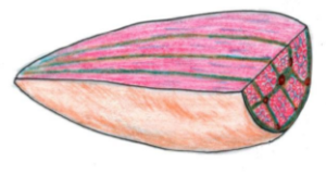
Fusiform – these are sometimes included within the parallel muscle group and are made up of spindle shaped fibres. A good example is the Biceps Brachii as the belly is wider than the origin and the insertion.
Muscle Movement
Muscles are only ever able to contract or pull. This means they have to work in groups and even when carrying out an action, do not work alone. A joint, therefore has to have two or more muscles working together. As a muscle contracts, the second muscle relaxes, and as this second muscle contracts, the first muscle relaxes. This is called Antagonistic action as they are pulling in the opposite direction to each other but without working against each other. One end of the muscle needs to be fixed, which is known as the origin and as that muscle contracts, the other end of the muscle moves towards the origin. The name given to the end of the muscle that moves towards the origin is called the insertion.
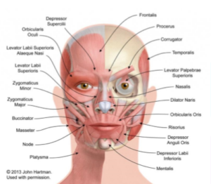
Frontalis Position Upper part of the cranium Action Elevates eyebrows; draws the scalp forwards Corrugator Position Inner corner of eyebrows Draws eyebrows together (frowning)
Procerus Position Top of nose between eyebrows Action Depresses the eyebrows (forms wrinkles over the nose)
Orbicularis Oculi Position Surrounds the eye, Action closes the eye (blinking)
Nasalis Position Over the front of nose Action Compresses nose (causing wrinkles)
Temporalis Position Runs downs the side of face towards jaw Action Aids chewing; closes mouth
Frontalis, Corrugator, Procerus, Orbicularis Oculi – are the muscles that we learn to treat in this Foundation Wrinkle reducing injection’s Module.